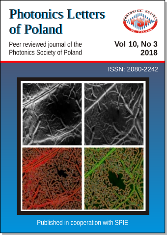
Enhancing microvasculature maps for Optical Coherence Tomography Angiography (OCT-A)
DOI:
https://doi.org/10.4302/plp.v10i3.841Abstract
OCT-A is becoming more popular in recent years and there is a high demand to improve the quality of angiograms as well as to extract quantitative information. We applied various processing methods for microvasculature enhancement like Hessian-Frangi to a data set obtained with Bessel and Gaussian OCT systems. We used angiogenesis, fractal and multifractal analysis to extract more quantitative information. We applied the processing methods for healthy, stroke, tumor progression and the results of enhanced processing and quantitative analysis for those are presented in this letter.Full Text: PDF
References
- J. G. Fujimoto, C. Pitris, S. A. Boppart, and M. E. Brezinski, "Optical coherence microscopy as a novel, non-invasive method for the 4D live imaging of early mammalian embryos", Neoplasia (New York, NY), 2000, 2(1-2):9-25 CrossRef
- O. Liba, E. D. SoRelle, D. Sen, and A. de la Zerda, "Contrast-enhanced optical coherence tomography with picomolar sensitivity for functional in vivo imaging", Sci Rep. 2016, 6(1):23337. CrossRef
- O Liba, M. D. Lew, E. D. SoRelle, et al., "Speckle-modulating optical coherence tomography in living mice and humans", Nat Commun. 2017, 8:15845. CrossRef
- K. Karnowski, A. Ajduk, B. Wieloch, et al., "Optical coherence microscopy as a novel, non-invasive method for the 4D live imaging of early mammalian embryos", Sci Rep. 2017, 7(1):4165. CrossRef
- V. J. Srinivasan, S. Sakadžić, I. Gorczynska, et al., "Quantitative cerebral blood flow with Optical Coherence Tomography", Opt Express. 2010, 18(3):2477. CrossRef
- S. Tamborski, H. C. Lyu, H. Dolezyczek, et al. Extended-focus optical coherence microscopy for high-resolution imaging of the murine brain. Biomed Opt Express. 2016, 7(11):4400-4414. CrossRef
- A. F. Frangi, W. J. Niessen, K. L. Vincken, and M. A. Viergever, "Multiscale vessel enhancement filtering", Lect Notes Comput Sc. 1998;1496:130–137 CrossRef
- A. A. Ucuzian, A. A. Gassman, A. T. East, and H. P. Greisler , Journal of burn care & research: official publication of the American Burn Association 2010 , 31(1):158. CrossRef
- R. Lopes, and Betrouni, N. (2009). "Fractal and multifractal analysis: A review", Med. Image Anal. 13, 634–649. CrossRef
- J. W. Baish and R. K. Jain., "Correspondence re: J. W. Baish and R. K. Jain, Fractals and Cancer. Cancer Res., 60: 3683-3688, 2000.", Cancer Res. 2000, Jul 15, 60(14):3683-8. DirectLink
Downloads
Published
2018-10-01
How to Cite
[1]
M. Rapolu, P. Niedźwiedziuk, D. Borycki, P. Wnuk, and M. Wojtkowski, “Enhancing microvasculature maps for Optical Coherence Tomography Angiography (OCT-A)”, Photonics Lett. Pol., vol. 10, no. 3, pp. 61–63, Oct. 2018.
Issue
Section
Articles


