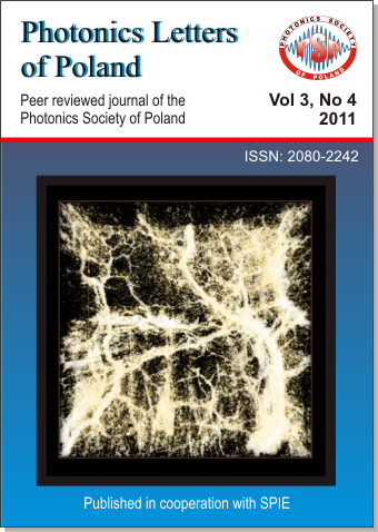
Imaging limbal and scleral vasculature using Swept Source Optical Coherence Tomography
DOI:
https://doi.org/10.4302/photon.%20lett.%20pl.v3i4.258Abstract
We demonstrate application of high-speed swept source optical coherence tomography for vessel visualization in the anterior segment of the human eye. The human corneo-scleral junction and sclera was imaged in vivo. Imaging was performed using a swept source OCT system operating at 1050nm wavelength range and 100kHz A-scan rate. The high imaging speed enables generation of 3D depth-resolved vasculature maps. The vessel visualization method revealed the rich vascular system in the conjunctiva and episclera.Full Text: PDF
References:
- D. Huang et al., "Optical coherence tomography", Science 254, 1178 (1991). [CrossRef]
- W. Drexler, J. G. Fujimoto (ed.), Optical Coherence Tomography. Technology and Applications (Springer, Berlin-Heidelberg 2008).
- A.M. Zysk, A. L. Oldenburg, D.L. Marks, F.T. Nguyen, S.A. Boppart, "Optical coherence tomography: a review of clinical development from bench to bedside", J. Biomed. Opt. 12, 051403 (2007). [CrossRef]
- Y. Wang, A. Fawzi, O. Tan, J. Gil-Flamer, D. Huang, "Retinal blood flow detection in diabetic patients by Doppler Fourier domain optical coherence tomography", Opt. Exp. 17, 4061 (2009). [CrossRef]
- M. Pircher, C.K. Hitzenberger, U. Schmidt-Erfurth, " Polarization sensitive optical coherence tomography in the human eye", Prog. Ret. Eye Res. 30, 431 (2011). [CrossRef]
- M. Wojtkowski, "High-speed optical coherence tomography: basics and applications", Appl. Opt. 49, D30 (2010). [CrossRef]
- J. A. Izatt et al., "Micrometer-Scale Resolution Imaging of the Anterior Eye In Vivo With Optical Coherence Tomography", Arch. Ophthalmol. 112, 1584 (1994). [CrossRef]
- M. Gora et al., "Ultra high-speed swept source OCT imaging of the anterior segment of human eye at 200 kHz with adjustable imaging range", Opt. Exp. 17, 14880 (2009). [CrossRef]
- B. Potsaid et al., "Ultrahigh speed 1050nm swept source / Fourier domain OCT retinal and anterior segment imaging at 100,000 to 400,000 axial scans per second", Opt. Exp. 18, 20029 (2010). [CrossRef]
- Y. Yasuno et al., "Three-dimensional and high-speed swept-source optical coherence tomography for in vivo investigation of human anterior eye segments", Opt. Exp. 13, 10652 (2005). [CrossRef]
- D.R. Anderson, Am. J. Ophthalmol. 108, 485 (1989).
- R.K. Wang, L. An, P. Francis, D.J. Wilson, "Doppler optical micro-angiography for volumetric imaging of vascular perfusion in vivo", Opt. Exp. 17, 8926 (2009). [CrossRef]
- B.J. Vakoc et al., "Three-dimensional microscopy of the tumor microenvironment in vivo using optical frequency domain imaging", Nature Med. 15, 1219 (2009). [CrossRef]
- K. Bizheva, N. Hutchins, L. Sorbara, A.A. Moayed, T. Simpson, "In vivo volumetric imaging of the human corneo-scleral limbus with spectral domain OCT", Biomed. Opt. Exp. 2, 1794 (2011). [CrossRef]
- L. Kagemann et al., "Identification and Assessment of Schlemm's Canal by Spectral-Domain Optical Coherence Tomography", Invest. Ophthalmol. Vis. Sci. 51, 4054 (2010). [CrossRef]
- L. Kagemann et al.,"3D visualization of aqueous humor outflow structures in-situ in humans", Exp. Eye Res., 93, 308 (2011). [CrossRef]
- P. Li et al., "In vivo microstructural and microvascular imaging of the human corneo-scleral limbus using optical coherence tomography", Biomed. Opt. Exp. 2, 3109 (2011). [CrossRef]
- M.A. Choma, M.V. Sarunic, Ch. Yang, J.A. Izatt, "Sensitivity advantage of swept source and Fourier domain optical coherence tomography", Opt. Exp. 11, 2183 (2003). [CrossRef]
Downloads
Additional Files
Published
2011-12-29
How to Cite
[1]
I. Grulkowski, J. J. Liu, B. Baumann, B. Potsaid, C. Lu, and J. G. Fujimoto, “Imaging limbal and scleral vasculature using Swept Source Optical Coherence Tomography”, Photonics Lett. Pol., vol. 3, no. 4, pp. pp. 132–134, Dec. 2011.
Issue
Section
Articles


